 Cherry tomatoes contaminated with salmonella: 92 sick and 1 dead
  A better coaching method can make a person grow
  What is the method to prevent diabetes in children?
  What are the effective factors in causing stomach ulcers?
  Why do embarrassing memories seem to appear at night?
  The amazing link between SARS-CoV-2 infection and newly started diabetes
  WHO says monkey pox is not a global emergency right now
  Single cell RNA sequencing uncovers new mechanisms of heart disease
  Hepatitis of unknown origin: 3 new deaths and 228 cases worldwide
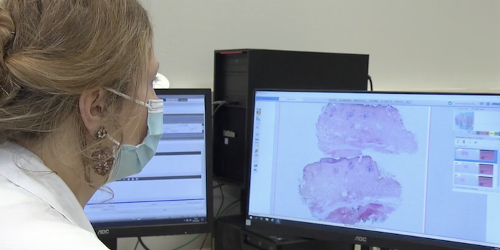
In pathological anatomy departments, white coats fix, cut and color samples from neighboring operating theaters. Tumors or potentially diseased organs. Until now, the microscope reigned supreme. But several months ago, at the Rennes University Hospital, it became a vestige, dethroned by a “high tech” scanner.
This device allows the slides to be digitized in very high definition and facilitates reading for the doctors responsible for the diagnosis. “It attracts the eye more quickly than with the microscope you can really see that the lesion is located here or here. While under the microscope it may be more difficult to find your way around, especially since you don’t have such a wide field, we will see a small part of the sample while there we have an overview “ explains Dr Solène Florence Kammerer-Jacquet, pathologist, at Rennes University Hospital.
Saving time for doctors
Another advantage: two clicks are enough to navigate in the patient’s file. Share it online too, to seek the expertise of other doctors. In this service, the switch to digital is a first step towards artificial intelligence. Tomorrow, it could make it even easier for doctors to do this.
“Artificial intelligence will help quantify cells, for example, will completely accelerate the reading of the slide, make it comfortable too, to free up medical time and so that the practitioner can devote himself to the most complex cases, wherever he is. has the most added value ” details Adrien Michaud, vice-president of APIDIM, the association for the innovation of medical devices. The Rennes University Hospital is the first establishment in France to be equipped with an anatomy and digital cytology department.